A Miracle of Survival: Overcoming Aortoesophageal Fistula in a 5-Year-Old
A 5-year-old boy arrived at our Emergency Room on the night of April 19th, after experiencing two episodes of hematemesis (vomiting blood) in the evening. He had been suffering from occasional abdominal pain for two weeks. Initially, he was taken to a local hospital for symptomatic treatment but arrived at our facility in severe distress, pale, and with low blood pressure.
Upon admission to our Pediatric Intensive Care Unit (PICU), the medical team promptly stabilized him with fluids, a blood transfusion, ionotropic support, and other necessary measures. Although initial tests revealed severe anaemia, no significant abnormalities were evident during systemic examination. An abdominal ultrasound (USG) showed normal results. The plan was to perform an endoscopy the next morning to further investigate.
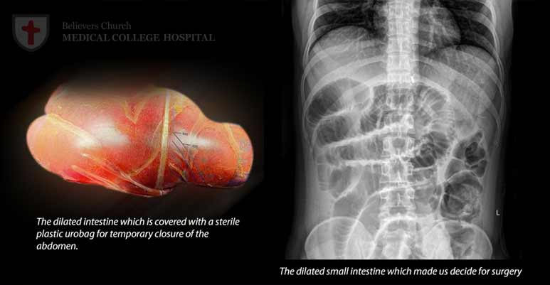
However, just a few hours after his admission, he had another massive episode of hematemesis, which led to his rapid deterioration. The medical team administered more fluids, increased ionotropic support, and initiated mechanical ventilation. A chest X-ray identified a coin-shaped foreign body (FB), which appeared to be lodged in the oesophagus. This discovery raised suspicion of button battery ingestion, prompting an immediate consultation with both Gastroenterology and Cardiovascular Surgery (CTVS) specialists.
Emergency esophagogastroduodenoscopy (OGD) confirmed the presence of a button battery embedded in the oesophagal wall with the erosion of the lateral oesophagal wall. Given that an oesophagal injury was unlikely to result in such severe bleeding, the medical team suspected an aortic injury and quickly arranged for an emergency CT aortogram, which confirmed an aortoesophageal fistula.
The child was rushed to the operating room for emergency thoracotomy and aortoesophageal repair. This procedure was high-risk, compounded by the challenge of securing enough O-negative blood for the transfusion.
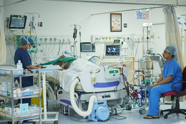
Post-surgery, his hemodynamics stabilized, but he developed multiple organ dysfunction syndrome (MODS) and acute kidney injury (AKI) as complications of severe hypotension, necessitating peritoneal dialysis. Additionally, the surgical team noted signs of infection in the oesophagal injury, leading to an upgrade in antibiotic treatment. Despite the use of Meropenam, the child continued to experience fever. Sterile cultures prompted a sepsis PCR panel, which tested positive for Enterobacteriaceae carrying the NDM and VIM genes.
In response, his antibiotics were further upgraded to Polymyxin B and, later, to Ceftazidime Avibactam plus Aztreonam due to his AKI. Over the next week, the child's condition gradually improved, and the fever subsided. Organ functions improved, peritoneal dialysis was discontinued, and he was extubated after five days.
During this period, a nasojejunal tube was inserted endoscopically, and the child was initiated on nasojejunal (NJ) feeds. All lines, drains, and catheters were eventually removed, and he seemed to be on a steady path to recovery. However, on the 12th night of his admission, he experienced another episode of hypotension and bleeding, necessitating another emergency thoracotomy. This time, the cause was chemical necrosis of the aorta and oesophagus. The necrotic aortic area was resected and repaired, and the oesophagus was reconstructed.
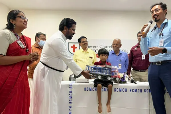
Following this second surgery, he was extubated after 24 hours, and feeds were reintroduced. The child, unfortunately, developed refeeding syndrome with severe electrolyte imbalances (hypokalemia, hypomagnesemia, and hypophosphatemia), which required management over several days. Feeds were gradually increased, and the child was progressively mobilized. On the 21st day after the second surgery, a contrast esophagogram showed no leaks, enabling the initiation of oral fluids on the 22nd day, which were then advanced to a soft diet and solid foods. The NJ tube was removed on the 35th day, and the child remained in the hospital for a total of 40 days.
An extensive literature review revealed the rarity of major vessel injuries in children, and data from the National Capital Poison Centre indicated that only a handful of individuals had survived aortoesophageal fistula cases between 1977 and 2019. This case represents a remarkable exception.
The successful outcome of this complex case can be attributed to the collaborative efforts of various medical specialities, including Pediatric ICU, Pediatrics, Nursing, CTVS, Gastroenterology, Nephrology, Anesthesia, Transfusion Medicine, PMR, and Dietetics. We are profoundly grateful to have played a role in this extraordinary journey of healing.
Other Stories
-

Going the Extra Mile: A Midnight…
" What does it truly mean to go the extra mile?
For Baby Hridaya Jith, it meant hope, healing, and…
-
Heartfelt Appreciation for Exceptional Care
I would like to express my heartfelt gratitude to the entire ICU team at your facility, with special mention to…
-
A Tale of Resilience: Suresh's Extraordinary…
Meet Mr. Suresh, a 27-year-old construction site supervisor with an inspiring story of overcoming adversity. Suresh's journey began when he…
Read more